![]() Index to Course Material
Index to Course Material
![]() Index to Section 10
Index to Section 10
![]() All Beta Folds
All Beta Folds
![]() Alpha/Beta Doubly Wound Folds
Alpha/Beta Doubly Wound Folds
You should be aware of alternative classifications, such as the Structural Classification of Proteins database (Alexey G. Murzin, Steven E. Brenner, Tim J.P. Hubbard, and Cyrus Chothia). In all, sixty-four categories of alpha/beta folds are listed. A number of the entries have links to diagrams by Manuel Peitsch.
With MAGE installed, study this Kinemage on Alpha Domain Structures, which accompanies the Branden and Tooze book.
Many enzymes are alpha/beta structures. Most alpha/beta proteins are cytosolic.
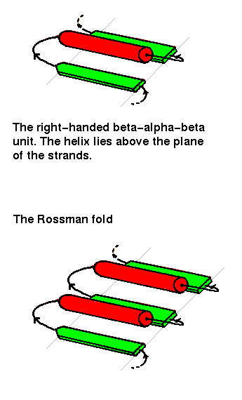
The beta-alpha-beta unit has already been described in a previous chapter . This motif is always right-handed. In alpha/beta structures, there is a repetition of this arrangement, giving a beta-alpha-beta-alpha.....etc sequence. The beta strands are parallel and hydrogen bonded to each other, while the alpha helices are all parallel to each other, and are anitparallel to the strands. Thus the helices form a layer packing against the sheet.
The beta-alpha-beta-alpha-beta subunit, often present in nucleotide-binding proteins, is named the Rossman Fold, after Michael Rossman (Rao and Rossman,1973).
Richardson (1981) names the alpha/beta structures "parallel alpha/beta domains", to denote the fact that each of the 2 secondary structures forms a parallel arrangement. Note that there is no obvious reason why one would not expect to find "parallel all alpha" (alpha-alpha-alpha subunit) folds, or "parallel all beta" (beta-beta-beta) folds in equally large numbers, but these do not occur. However, the marked tendency for helices to pack aligned with sheets has been explained by the "complementary twist" model (Chothia et al. , 1977; see Section 9).
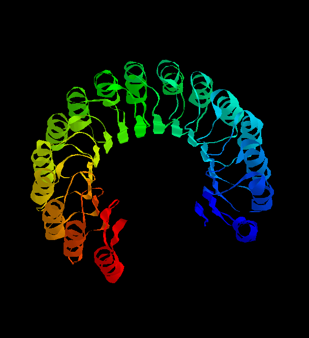 The
structure of the remarkable placental ribonuclease inhibitor
(Kobe, B. & Diesenhofer, J. (1993 ) Nature V.366, 751- )takes the
concept of the repeating alpha/beta unit to extremes. It is a cytosolic
protein that binds extremely strongly to any ribonuclease that may leak into the
cytosol.
The
structure of the remarkable placental ribonuclease inhibitor
(Kobe, B. & Diesenhofer, J. (1993 ) Nature V.366, 751- )takes the
concept of the repeating alpha/beta unit to extremes. It is a cytosolic
protein that binds extremely strongly to any ribonuclease that may leak into the
cytosol.
![]() Take a look 1bnh (286Kb)
[Bbk|BNL|ExP|Waw|Hal]
...28Kb GIF
and you'll see the 17-stranded parallel beta sheet curved into an open
horseshoe shape, with 16 alpha-helices packed against the outer surface.
It doesn't form a barrel although it looks as though it should. The strands are only
very slightly slanted, being nearly parallel to the central `axis'.
Take a look 1bnh (286Kb)
[Bbk|BNL|ExP|Waw|Hal]
...28Kb GIF
and you'll see the 17-stranded parallel beta sheet curved into an open
horseshoe shape, with 16 alpha-helices packed against the outer surface.
It doesn't form a barrel although it looks as though it should. The strands are only
very slightly slanted, being nearly parallel to the central `axis'.
If the first strand hydrogen
bonds to the last, then the structure closes on itself forming a barrel-like
structure. This is shown in the
picture of triose phosphate isomerase (35 Kb):

![]() 1tim (322Kb)
[Bbk|BNL|ExP|Waw|Hal]
... SCRIPT
1tim (322Kb)
[Bbk|BNL|ExP|Waw|Hal]
... SCRIPT
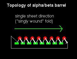
Note that the "staves" of the barrel are slanted, due to the twist of the beta sheet. Also notice that there are effectively four layers to this structure. The direction of the sheet does not change (it is anticlockwise in the diagram). Such a structure may therefore be described as singly wound.
The packing interactions of the core residues of alpha/beta barrels is described in Section 9.
Doubly Wound Alpha/Beta Topologies
In a structure which is open rather than closed like the barrel, helices would
be situated on only one side of the beta sheet if the sheet direction did not
reverse (see previous figure.
). Therefore open alpha/beta structures must be
doubly wound to cover both sides of the sheet.
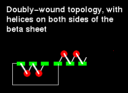
The chain starts in the middle of the sheet and travels outwards, then returns to the centre via a loop and travels outwards to the opposite edge:
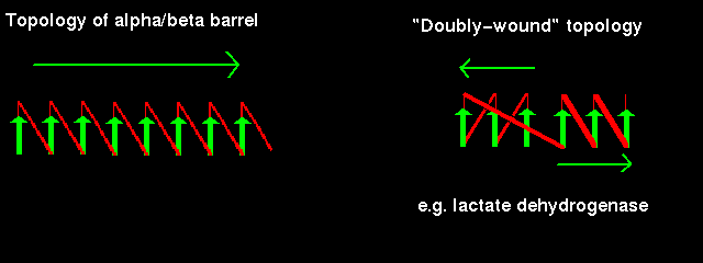
Doubly-wound topologies where the sheet begins at the edge and works inwards are rarely observed.
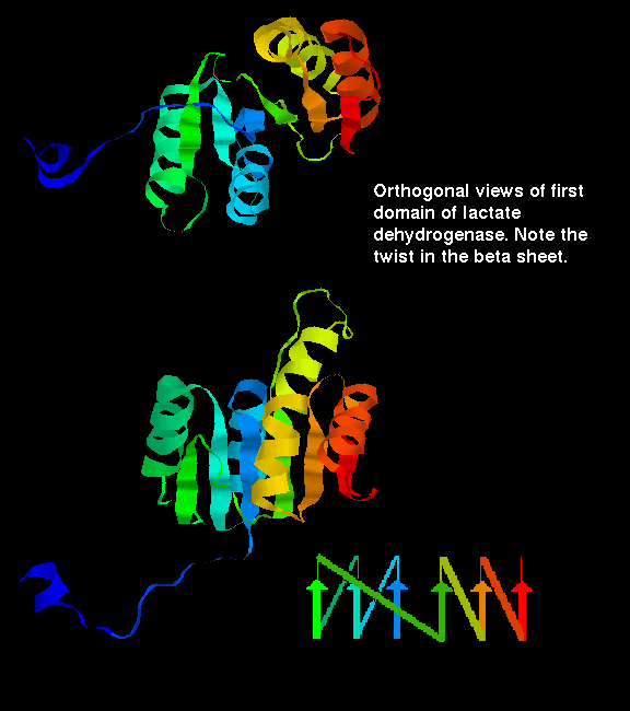
Click here for a diagram illustrating the doubly wound fold of the first domain of lactate dehydrogenase.
![]() or examine the crystal structure 9ldb (468Kb)
[Bbk|BNL|ExP|Waw|Hal]
... SCRIPT to display the N-terminal domain of chain
A as in the diagram
or examine the crystal structure 9ldb (468Kb)
[Bbk|BNL|ExP|Waw|Hal]
... SCRIPT to display the N-terminal domain of chain
A as in the diagram
Last updated 8th Aug '96