 All-Beta Topologies
 Index to Course Material
 Index to Section 10
 All Alpha Folds
 Alpha/Beta Folds
All-Beta Topologies
Introduction
Again, this is a review of some of the 'classic' examples.
You should also browse through the
Mainly Beta Class of the
CATH Protein
Structure Classification Database at University College, London. This
lists 7 different architectures.
You should also be aware of alternative classifications, such as
the
Structural Classification of Proteins database
(Alexey G. Murzin, Steven E. Brenner, Tim J.P. Hubbard, and Cyrus Chothia). In all, fifty-two
categories of all beta folds are listed.
A number of the entries have links to diagrams by Manuel Peitsch.
With MAGE installed, study this
Kinemage
on Alpha Domain Structures, which accompanies the
Branden and Tooze book.
Protein folds which consist of almost entirely beta sheets
exhibit a completely or mostly antiparallel arrangement. Many of these antiparallel domains
consist of two sheets packed against each other, with
hydrophobic side chains forming the interface
(refer to Section 9). Bearing in mind that side chains
of a beta-strand point alternately to opposite sides of a sheet, this means
that such structures will tend to have a sequence of alternating hydrophobic and polar
residues.
Beta Sandwiches and Beta Barrels
The immunoglobulin fold is introduced in the section on
mosaic proteins . In this fold,
the strands form two sheets packed against each other, forming a "beta
sandwich". Also look at the
fibronectin type 3 domain in the same section.
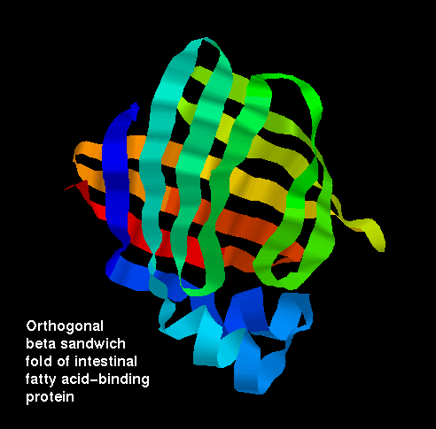
Aligned and Orthogonal Beta Sandwiches
The packing arrangements in both aligned and orthogonal beta-sandwiches has
been described in Section 9.
 Click here for a diagram
of this beta sheet arrangement in
the lipocalin intestinal fatty acid-binding protein.
Click here for a diagram
of this beta sheet arrangement in
the lipocalin intestinal fatty acid-binding protein.
 1ifb (95Kb)
[Bbk|BNL|ExP|Waw|Hal]
... SCRIPT
1ifb (95Kb)
[Bbk|BNL|ExP|Waw|Hal]
... SCRIPT
This is just one member of the
Lipocalin family, which bind small
molecules between the sheets of the sandwich.
Beta barrels
Some antiparallel beta sheet domains are better described as beta
barrels rather than beta sandwiches,
for example streptavadin.
1stp (87Kb)
[Bbk|BNL|ExP|Waw|Hal]
See also
the diagrams of porin in the section on
membrane proteins in the previous chapter. Note that some structures are
intermediate between the extreme barrel and sandwich arrangements.
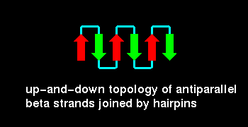
Up-and-down Antiparallel Beta Sheets
The simplest topology for an antiparallel beta sheet involves
loops connecting adjacent strands, as shown:
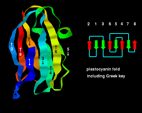
The Greek Key Topology
The Greek Key topology, named after a pattern that was
common on Greek pottery, is shown below. Three up-and-down beta
strands connected by hairpins are followed by a longer connection
to the fourth strand, which lies adjacent to the first.
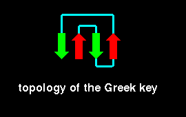
Folds including the Greek key topology have been found to have
5-13 strands. An example is given below.
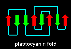 plastocyanin (21Kb GIF).
2plt (71Kb)
[Bbk|BNL|ExP|Waw|Hal]
Here is the crystal
structure. Notice that this has a mixed sheet- there are two parallel
pairs of strands. This SCRIPT renders the molecule
as in the diagram.
plastocyanin (21Kb GIF).
2plt (71Kb)
[Bbk|BNL|ExP|Waw|Hal]
Here is the crystal
structure. Notice that this has a mixed sheet- there are two parallel
pairs of strands. This SCRIPT renders the molecule
as in the diagram.
Gamma-crystallin
Gamma-crystallin has two domains each of which is an eight-
stranded beta barrel-type structure composed of two Greek
keys:
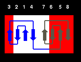
In fact, the structure is more accurately described as consisting
of two beta sheets, one consisting of strands 2,1,4,7 (white) and the
other of strands 6,5,8,3 (red) as indicated in the diagram.
This can be seen by examining the crystal structure of gamma-crystallin.
2gcr (136Kb)
[Bbk|BNL|ExP|Waw|Hal]
This SCRIPT displays only the N-terminal domain, and
colours the two sheets as in the diagram above (white and red). To distinguish the
two Greek keys, type the following:
select 1-39
colour blue
select 40-80
colour [90,90,70]
Sequence homology has been found between the
two Greek key motifs within each domain, and also between the two domains
themselves. The latter homology is higher than the former; this implies that the
structure evolved from a single Greek key fold by means of a gene duplication to
produce a domain of two Greek keys, followed by a second duplication resulting in
two similar domains. This is supported by the fact that in some crystallins each
Greek key motif is coded by a different exon, with introns between them.
The Jellyroll Topology
Richardson(1981) describes the jellyroll fold as being
formed by the addition of an extra "swirl" to a Greek key:
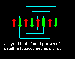 Click here for a diagram
illustrating this fold in the coat protein of
satellite tobacco necrosis virus (21Kb GIF).
Click here for a diagram
illustrating this fold in the coat protein of
satellite tobacco necrosis virus (21Kb GIF).
2stv (158Kb)
[Bbk|BNL|ExP|Waw|Hal]
... SCRIPT to render as in
the diagram
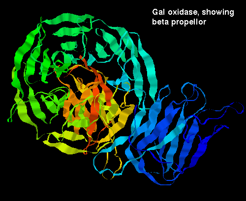
Beta Propellors
 2bat (304Kb)
[Bbk|BNL|ExP|Waw|Hal]
Click here to examine the crystal structure of
one subunit (about 470 residues) of neuraminidase. One molecule is
composed of four of these subunits. Each is a superbarrel of six
four-stranded antiparallel sheets. The whole structure has a basically
up-down topology as shown:
2bat (304Kb)
[Bbk|BNL|ExP|Waw|Hal]
Click here to examine the crystal structure of
one subunit (about 470 residues) of neuraminidase. One molecule is
composed of four of these subunits. Each is a superbarrel of six
four-stranded antiparallel sheets. The whole structure has a basically
up-down topology as shown:
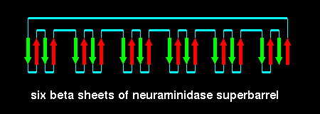
This fold is called a beta propellor; there are six 'blades' in
neuraminidase.
Here are two other examples - how many blades are there in each propellor?:
Beta Trefoils
This fold has an approximately 3-fold axis of symmetry.
Examine the crystal
structure of erythrina trypsin inhibitor 1tie (114Kb)
[Bbk|BNL|ExP|Waw|Hal]
... SCRIPT
"Beta Helix"
This dramatically unusual fold was discovered quite recently. The beta strands
wind round the structure describing a helical topology, as can be seen in this
image of pectate lysase.
2pec (245Kb)
[Bbk|BNL|ExP|Waw|Hal]
Index to Course Material
Index to Section 10
All Alpha Folds
Alpha/Beta Folds
Last updated 26th Jun '96
 Click here for a diagram
of this beta sheet arrangement in
the lipocalin intestinal fatty acid-binding protein.
Click here for a diagram
of this beta sheet arrangement in
the lipocalin intestinal fatty acid-binding protein.
 plastocyanin (21Kb GIF).
 Click here for a diagram
illustrating this fold in the coat protein of
satellite tobacco necrosis virus (21Kb GIF).
Click here for a diagram
illustrating this fold in the coat protein of
satellite tobacco necrosis virus (21Kb GIF).