|
|
|
Quaternary Structure - Multimeric Enzymes
This page describes some further examples of multimeric enzymes (some others
are described in the overview of this Section.
Enzymes are biological catalysts, which mediate a vast number of reactions
in the biochemical pathways of life. Apart from some RNA molecules which
have been relatively recently shown to have catalytic activity (ribozymes),
all enzymes are proteins. They are highly specific in the reactions they
catalyse, and bind their substrates by non-bonded interactions (refer to
the appropriate page in Section 7 of
the course). The catalytic regions of the enzymes which bind the substrates
are called active sites. In some cases, oligomerization is a requisite
for the existence of a complete active site; make sure you have read the
overview of this Section, which describes two
examples.
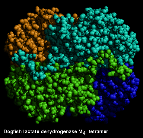
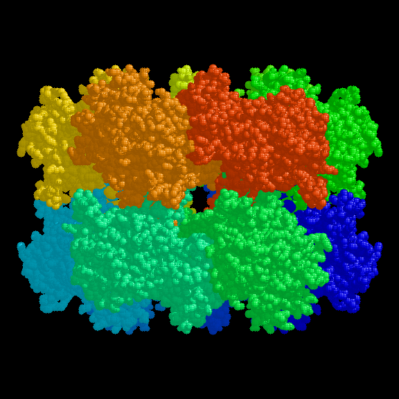
This is a tetrameric enzyme. In certain species there are two forms of the subunit, H and M, which results in a family of different isozymes. In heart muscle, the H4 form predominates, while M4 is the major form in skeletal muscle. Even though the two chains are significantly different, the MH3, M2H2 and M3H forms are found in the expected quantities, as the interface regions of both types are similar.
Consider how many distinct forms of M2H2 are possible.
The catalytic function does not involve interactions between the subunits, as the kinetic behaviour of, for example, the H3M form is the same as a 3:1 mixture of H4 and M4.
![]() Examine the
monomer 1ldm (260Kb)
[Bbk|BNL|ExP|Waw|Hal].
Examine the
monomer 1ldm (260Kb)
[Bbk|BNL|ExP|Waw|Hal].
 This diagram shows the four
subunits.
This diagram shows the four
subunits.
Here is the tetramer of the M4 isozyme, courtesy of Brookhaven PDB.
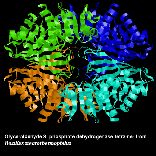
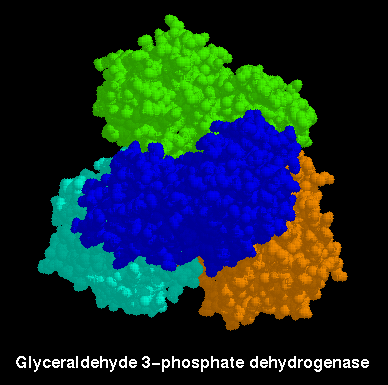
Note the similarity between the arrangements of this enzyme and lactate dehydrogenase. What type of symmetry is this? Make sure you can identify the axes of symmetry.
![]() In this case
however, the asymmetric unit of the crystal structure includes the complete
tetramer 1gd1 (966Kb)
[Bbk|BNL|ExP|Waw|Hal].
In this case
however, the asymmetric unit of the crystal structure includes the complete
tetramer 1gd1 (966Kb)
[Bbk|BNL|ExP|Waw|Hal].
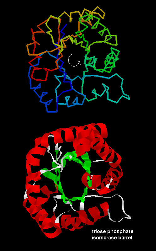
The alpha/beta barrel structure of this enzyme has been described in the previous chapter . Here is a diagram .
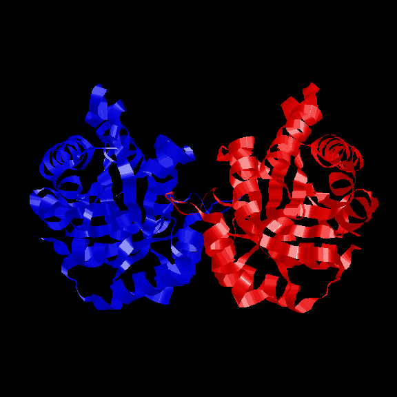
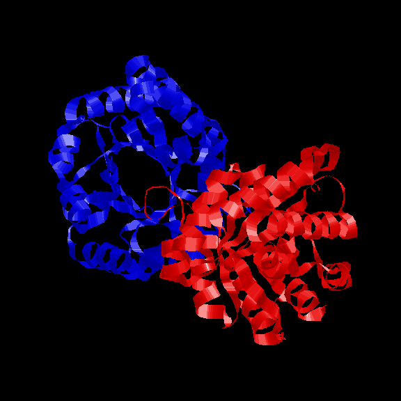
Like many other structures of this fold, such as some of the other glycolytic enzymes, this enzyme is dimeric. However, there is no evidence for cooperativity of substrate binding during catalysis. Here are two diagrams indicating the symmetrical arrangement of the 2 subunits and the relative orientations of the 2 barrel axes .
![]() Examine the
crystal structure 1tim (322Kb)
[Bbk|BNL|ExP|Waw|Hal].
Examine the
crystal structure 1tim (322Kb)
[Bbk|BNL|ExP|Waw|Hal].
The biologically active form of this enzyme is a dodecamer. The structure consists of two hexagonal rings, with an aqueous channel through the middle, stacked against each other.
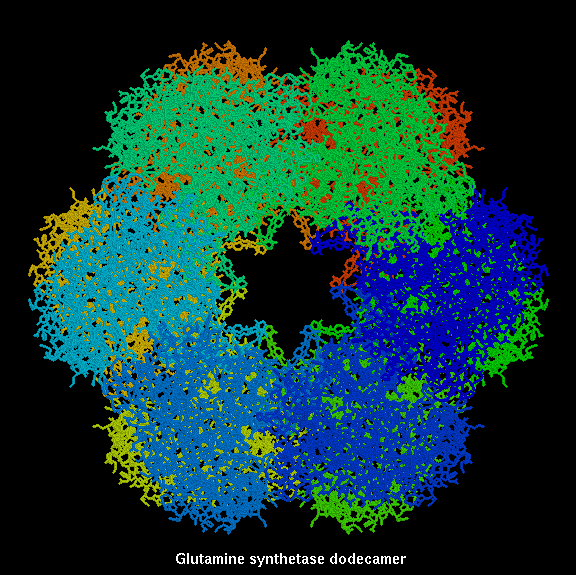
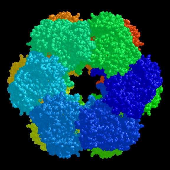
Click here for a diagram of a single chain, courtesy of The Swiss-3DImage Collection , who also provide several further views of the dodecamer ; and also detail of the active site .
![]() Here is the
crystal structure (the asymmetric unit is an entire dodecamer) 2gls
(3.6Mb)
[Bbk|BNL|ExP|Waw|Hal].
Here is the
crystal structure (the asymmetric unit is an entire dodecamer) 2gls
(3.6Mb)
[Bbk|BNL|ExP|Waw|Hal].
As always, don't forget that there are numerous references cited within the Protein Data Bank files which are pointed to by the hypertext.
|
|
|
Last updated 14th April '97