Last modified 19th April '95 © Birkbeck College 1995
Back to main PPS Index
Back to Quaternary Structure Index
Examples of Multimeric Enzymes
Dehydrogenases
Various dehydrogenases have been introduced in the section on
enzymes in the chapter on
tertiary structure .
Alcohol Dehydrogenase
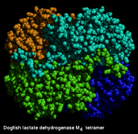
Lactate Dehydrogenase
This is a tetrameric enzyme. In certain species there are two forms of the
subunit, H and M, which results in a family of different
isozymes. In heart muscle, the H4 form predominates, while M4 is the major
form in skeletal muscle. Even though the two chains are significantly
different, the MH3, M2H2 and M3H forms are found in the expected quantities,
as the interface regions of both types are similar.
Consider how many distinct forms of M2H2 are possible.
The catalytic function does not involve interactions between the subunits, as
the kinetic behaviour of, for example, the H3M form is the same as a 3:1 mixture
of H4 and M4.
Examine the monomer .
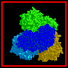
 This diagram shows the four subunits.
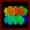
Here is the tetramer of the M4 isozyme, courtesy of
BROOKHAVEN.
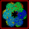
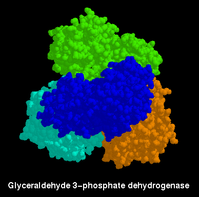
Glyceraldehyde 3-Phosphate Dehydrogenase
Note the similarity between this enzyme and lactate dehydrogenase:

Here is the complete
tetramer .
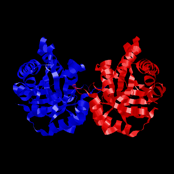
Triose Phosphate Isomerase
The alpha/beta barrel structure of this enzyme has been described in the
previous chapter . Here is a
diagram .
Like many other structures of this fold, such as some of the
glycolytic enzymes , this
enzyme is dimeric. Here are two diagrams indicating
the symmetrical arrangement of the 2 subunits and
the relative orientations of the 2 barrel axes .
Examine the crystal
structure .
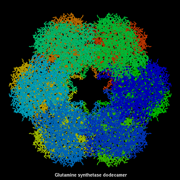
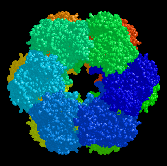
Glutamine Synthetase
The biologically active form of this enzyme is a dodecamer. The structure consists
of two hexagonal rings, with an aqueous channel through the middle, stacked
against each other.

Click
here for a diagram of a single chain, courtesy of
The Swiss-3DImage Collection
, who also provide
several further
views of the
dodecamer ; and also detail of the
active site .
Here is the crystal
structure .
References
- Abad-Zapatero, C., Griffith, J.P., Sussman, J.L. and Rossmann, M.G. (1987)
Refined crystal structure of dogfish M4 apo-lactate dehydrogenase,
J. Mol. Biol. 198, 445
- Banner, D.W., Bloomer, A.C., Petsko, G.A., Phillips, D.C. and Wilson,
I.A. (1976) Atomic coordinates for triose phosphate isomerase from chicken
muscle, Biochem. Biophys. Res. Comm. 72, 146
- Eklund, H., Samama, J.-P., Wallen, L., Branden, C.-I., Akeson, A. and
Jones, T.A. (1981) Structure of triclinic ternary complex of horse liver
alcohol dehydrogenase at 2.9Å resolution, J. Mol. Biol. 146,
561
- Fersht, A.R. (1985) Enzyme Structure and Mechanism, W.H. Freeman & Co.,
New York, pp. 390-404
- Skarzynski, T., Moody, P.C.E. and Wonacott, A.J. (1987) Structure of
holo-glyceraldehyde-3-phosphate dehydrogenase from Bacillus
stearothermophilus at 1.8Å resolution, J. Mol. Biol. 193
,171
- Yamashita, M.M., Almassy, R.J., Janson, C.A., Cascio, D. and
Eisenberg, D. (1989) Refined atomic model of glutamine synthetase at 3.5Å
resolution J. Biol. Chem. 264, 17681
As always, don't forget that there are numerous references cited within the
Protein Data Bank files which are pointed to by the hypertext.
Back to Main PPS Index
J. Walshaw

 This diagram shows the four subunits.

