3.0 Types of Secondary Structure
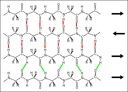
Beta sheets are found in two forms designated as "Antiparallel" or "Parallel" based on the relative directions of two interacting beta strands. The average length of a beta sheet is about 6 residues and most beta sheets contain less than 6 strands. Side chains from adjacent residues of a strand in a beta sheet are found on opposite sides of the sheet and do not interact with one another. Therefore, like alpha-helices, beta-sheets have the potential for amphiphilicity with one face polar and the other apolar. However, unlike alpha-helices which are comprised of residues from a continuous polypeptide segment (i.e., hydrogen bonds between CO of residue i and NH of residue i+3), beta sheets are formed from strands that are very often from distant portions of the polypeptide sequence. Hydrogen bonds in beta sheets are on average 0.1 Angstrom shorter than those found in alpha helices (Baker & Hubbard, 1984).
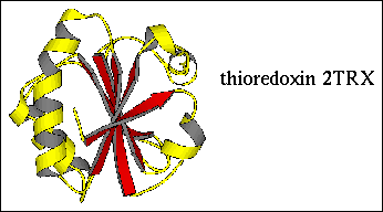
Figure 8. The protein thioredoxin (2TRX.PDB) contains a five-stranded beta sheet comprised of three parallel strands and three antiparallel strands. The entire protein is shown as a cartoon with the beta strands (three parallel strands and three antiparallel strands) colored red and alpha helices colored yellow.
Half of the backbone hydrogen bond donors and acceptors in strands at the edges of a beta sheets are uninvolved in the sheet interactions. Unlike the situation with alpha helices ("helix capping" 3.1.4.1"), capping residues in beta sheets have not yet been characterized (interesting project??). Proline is sometimes found in antiparallel beta sheets and in all cases I am aware of, the imide nitrogen points away from the strand interior. A good example of this is found in dendrotoxin K (1DTK; Berndt et al., 1993), a BPTI homologue which has a proline in each strand of the beta hairpin. Sheets can fold onto themselves forming a barrel or cylinder, thus eliminating the non-hydrogen bonded "ends" (see the tim barrel in Figure 1 ).
Generated with CERN WebMaker