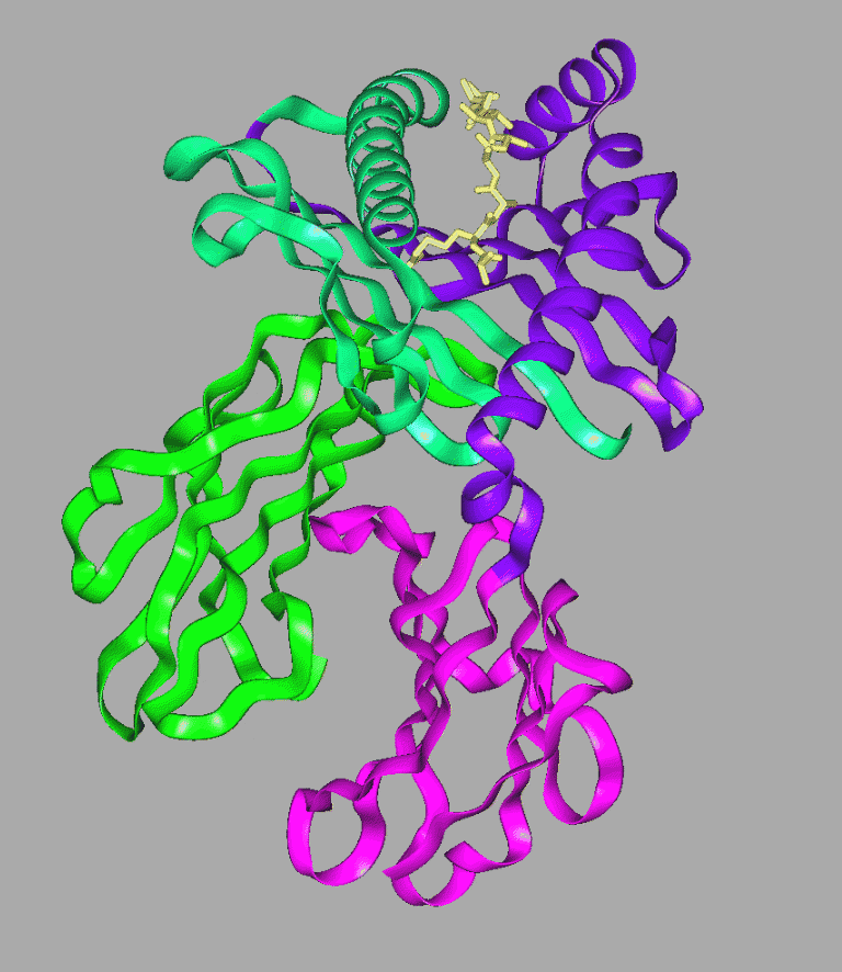
MHC Class I is a membrane spanning molecule composed of two proteins. The membrane spanning protein is approximately 350 amino acids in length, with about 75 amino acids at the carboxylic end comprising the transmembrane and cytoplasmic portions. The remaining 270 amino acids, as shown in the ribbon diagram, are divided into three globular domains labelled Alpha-1 (blue-green), Alpha-2 (purple) and Alpha-3 prime (magenta), with alpha-1 being closest to the amino terminus and alpha-3 closest to the membrane. The second portion of the molecule is a small globular protein called Beta-2 Microglobulin (green) It associates primarily with the alpha-3 prime domain and is necessary for MHC stability. The bound peptide (cream) sits within the groove.
The three-dimensional structure of the Class I Human Histocompatibility Protein, HLA-B27 has been determined at 2.1 angstroms resolution. If your Web Browser has been suitably configured, download this crystal structure of Human MHC Class I, [1HSA] (563Kb) - this is a dimer [Bbk|BNL|Hal]
The MHC molecules ability to present a wide range of antigenic peptides for T cell recognition requires a compromise between broad specificity and high affinity. Three-dimensional structures of both class I and class II MHC molecules show a unique structural solution to this problem. The peptide main chain is tightly bound whilst peptide side chains show less restrictive interactions. It is primarily the peptide side-chain contacts and conformational variability that ensures that the peptide-MHC complex presents an antigenically unique surface to T cell receptors.
The class I histocompatibility antigen from human cell membranes has two structural motifs: the end furthest from the membrane contains two domains with immunoglobulin-folds and the distal end has a platform of eight antiparallel beta-strands topped by alpha-helices. Superposition of the structurally similar domains found in the heterodimer show differences mainly in the turn regions. A large groove between the alpha-helices provides a binding site for processed foreign antigens. Examination of this groove shows that the solvent-accessible amino acids along the centre and widest end of the cleft are the most polymorphic in mouse and human alleles . Conserved side-chains are clustered at the narrower ends of the groove. Six pockets in the antigen-binding groove, of variable shape and composition, bind side-chains from antigenic peptides.
Maffei A, 1997
MHC class I antigen processing pathways.
Hum Immunol 54(2), 91-103 (1997)
OMIM
*142800
HLA-A HISTOCOMPATIBILITY TYPE; HLAA
References for MHC Class I Peptide Binding Motifs