Last modified 19th May '95 © Birkbeck College 1995
Back to main PPS Index
Back to Protein Interactions Index
Glycosidases
These enzymes cleave oligosaccharides by hydrolysis.
Some examples:
Cellulases
Various species of fungi produce enzymes which hydrolyze cellulose.
Trichoderma reesei produces two endoglucanases, endoglucanase I (EG1)
and endoglucanase II (EGII) and two exoglucanases,
cellobiohydrolase I (CBHI) and cellobiohydrolase II (CBHII).
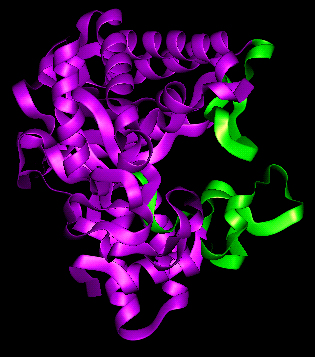
All four of these enzymes contain a highly homologous 36-residue region called
the A domain
(here is a
picture from The
Swiss-3DImage Collection), connected
to the (enzymatically active) core domain by a threonine- and serine-rich
linking sequence. The A domain has no catalytic activity in CBHI and CBHII but
is thought to have a cellulose-binding function, as the core protein alone
does not have full cellulose-hydrolyzing activity (but has normal activity on small
synthetic substrates). In EGI and CBHI the A domain is at the C-terminus, while
it is at the N-terminus in EGII and CBHII.
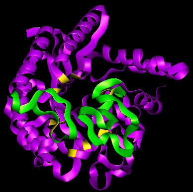
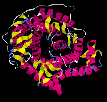
CBHI and CBHII produce cellobiose (a disaccharide). The substrate of CBHII
requires at least three consecutive ß(1-4)-bonded glycosidic units for the
enzyme to cleave a glycosidic bond. The proposed active site has four subsites
(Rouvinen et al., 1990).
Two extensive loops (residues 172-189, 394-429) occur at the C-terminal end
of the alpha/beta barrel, which form an enclosed tunnel approximately 20Å
in length, in which the substrate is believed to be bound.
Here is a view down this tunnel (the loop residues
mentioned above are coloured green) and a view down the
axis of the alpha/beta barrel, in which the positions of some of the more
important residues of the active site are coloured yellow. Examine the
pdb structure file
(C-alpha atoms only).
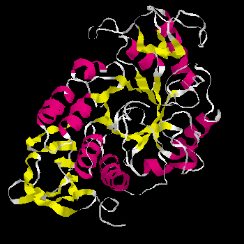
Alpha-amylase (Taka Amylase A)
 pdb file
 diagram
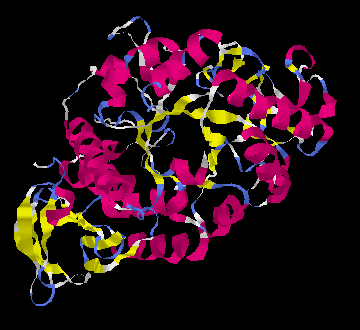
Acid alpha-amylase
pdb file
diagram
Beta-amylase
pdb file
Glycosidases with other folds
Pectate Lyase
pdb file
diagram
Glucoamylase [check this- what fold is it?]
pdb file
diagram
Neuraminidase
pdb file
refer to this section
in the Chapter on Protein Folds
Xylanase
pdb file
diagram 1
diagram 2
pdb file
Click here for the page on lysozyme.
References
- Aleshin, A., Golubev, A., Firsov, L.M. and Honzatko, R.B. (1992) Crystal
structure of glucoamylase from
Aspergillus awamori var. X100 to 2.2Å resolution J. Biol.
Chem. 267, 19291
- Boel, E., Brady, L., Brzozowski, A.M., Derewenda, Z., Dodson, G.G.,Jensen,
V.J., Petersen, S.B., Swift, H., Thim, L., and Woldike, H.F. (1990) Calcium
binding in alpha-amylases: an X-ray diffraction study at 2.1Å resolution of
two enzymes from Aspergillus Biochemistry, 29 6244
- Campbell, R.L., Rose, D.R., Wakarchuk, W.W., To, R.J., Sung, W. and Yaguchi,
M. High-resolution structures of xylanases from B. circulans and
T. harzianum identify a new folding pattern and implications for the
atomic basis of the catalysis, to be published
- Harris, E., Aleshin, A., Firsov, L. and Honzatko, R.B. (1993) Refined
structure for the complex of 1-deoxynojirimycin with glucoamylase from
Aspergillus awamori var. X100 to 2.4Å resolution Biochemistry
32, 1618
- Matsuura, Y., Kusunoki, M., Harada, W. and Kakudo, M. (1984) Structure and
possible catalytic residues of taka-amylase A J. Biochem. (Tokyo) 95, 697
- Matsuura, Y., Kusunoki, M., Harada, W., Tanaka, N., Iga, Y., Yasuoka, N.,
Toda, H., Narita, K. and Kakudo, M. (1980) Molecular structure of taka-amylase A
I. Backbone chain folding at 3Å resolution J. Biochem. (Tokyo)
87, 1980
- Mikami, B., Sato, M, Shibata, T., Hirose, M., Aibara, S., Katsube, Y. and
Morita, Y. (1992) Three-dimensional structure of soybean beta-amylase determined
at 3.0Å resolution: preliminary chain tracing of the complex with
alpha-cyclodextrin J. Biochem. (Tokyo) 112, 541
- Rouvinen, J., Bergfors, T., Teeri, Y., Knowles, J.K.C. and Jones, T.A.
(1990) Three-dimensional structure of Cellobiohydrolase Science 249
, 380-386
- Varghese, J.N. and Colman, P.M. (1991) Three-dimensional structure of the
neuraminidase of influenza virus A/Tokyo/3/67 at 2.2Å resolution
J. Mol. Biol. 473
- Yoder, M.D., Keen, N.T. and Jurnak, F. (1993) New domain motif: the structure
of pectate lyase C, a secreted plant virulence factor Science 260,
1503
- Yoder, M.D., Lietzke, S.E. and Jurnak, F. (1993) Unusual structural features
of the parallel beta-helix of the pectate lyases Structure (London)
1, 241
Refer to the
page
on glycosidases in the
Enzyme Structures Database at UCL, and also the
glyosidases page of the
Enzyme nomenclature database at ExPASy .
Kinemages
Here is the index to the Protein Science
Kinemages on Glycosidases
.

Back to Main PPS Index
J. Walshaw
 diagram
 diagram
 diagram
 diagram
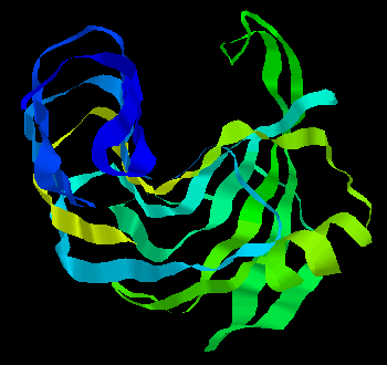 diagram 1
 diagram 2