Last modified 16th May '95 © Birkbeck College 1995
Back to main PPS Index
Back to Protein Interactions Index
Glycosidases
These enzymes cleave oligosaccharides by hydrolysis (of which bonds in
particular?).
Some examples:
Cellulases(?)
Various species of fungi produce enzymes which hydrolyze cellulose.
Trichoderma reesei produces two endoglucanases, endoglucanase I (EG1)
and endoglucanase II (EGII) and two exoglucanases,
cellobiohydrolase I (CBHI) and cellobiohydrolase II (CBHII).
All four of these enzymes contain a highly homologous 36-residue region called
the A domain
(here is a
picture from The
Swiss-3DImage Collection), connected
to the (enzymatically active) core domain by a threonine- and serine-rich
linking sequence. The A domain has no catalytic activity in CBHI and CBHII but
is thought to have a cellulose-binding function, as the core protein alone
does not have full activity on cellulose (but has normal activity on small
synthetic substrates). In EGI and CBHI the A domain is at the C-terminus, while
it is at the N-terminus in EGII and CBHII.
CBHI and CBHII produce cellobiose (a disaccharide). The substrate of CBHII
requires at least three consecutive ß(1-4)-bonded glycosidic units for the
enzyme to cleave a glycosidic bond. The proposed active site has four subsites
(Rouvinen et al., 1990).
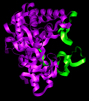
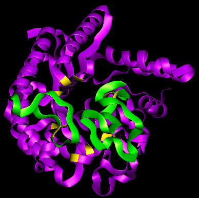
Two extensive loops (residues 172-189, 394-429) occur at the C-terminal end
of the alpha/beta barrel, which form an enclosed tunnel approximately 20Å
in length, in which the substrate is believed to be bound.
Here is a view down this tunnel (the loop residues
mentioned above are coloured green) and a view down the
axis of the alpha/beta barrel, in which the positions of some of the more
important residues of the active site are coloured yellow. Examine the
pdb structure file
(C-alpha atoms only).
Glucoamylase [check this- what fold is it?]
 pdb structure file
Alpha-amylase (Taka Amylase A)
pdb file
diagram
Acid alpha-amylase
pdb file
diagram
Beta-amylase
pdb file
Glycosidases with other folds
Neuraminidase
pdb file
refer to this section
in the Chapter on Protein Folds
Xylanase
pdb file
diagram 1 diagram 2
Lysozyme
pdb file
Click here for the page on lysozyme.
References
- Rouvinen, J., Bergfors, T., Teeri, Y., Knowles, J.K.C. and Jones, T.A.
(1990) Three-dimensional structure of Cellobiohydrolase Science 249
, 380-386
-
Refer to the
page
on glycosidases in the
Enzyme Structures Database at UCL, and also the
glyosidases page of the
Enzyme nomenclature database at ExPASy .
Kinemages
Here is the index to the Protein Science
Kinemages on Glycosidases
.
Back to Main PPS Index
J. Walshaw
