 Page 1 (in Ch.7)
Page 2 Tertiary
Structure; Substrates
Page 3 Active
Site
Page 1 (in Ch.7)
Page 2 Tertiary
Structure; Substrates
Page 3 Active
Site
 Page 5 Reaction
Mechanism
Page 6 Crystal
Structures
Page 7 References
Page 5 Reaction
Mechanism
Page 6 Crystal
Structures
Page 7 References
Back to Protein Interactions Index
Page 1 (in Ch.7)
Page 2 Tertiary
Structure; Substrates
Page 3 Active
Site
Page 5 Reaction
Mechanism
Page 6 Crystal
Structures
Page 7 References

in loop
between colour in
domain strands: diagrams
______ ________ _________
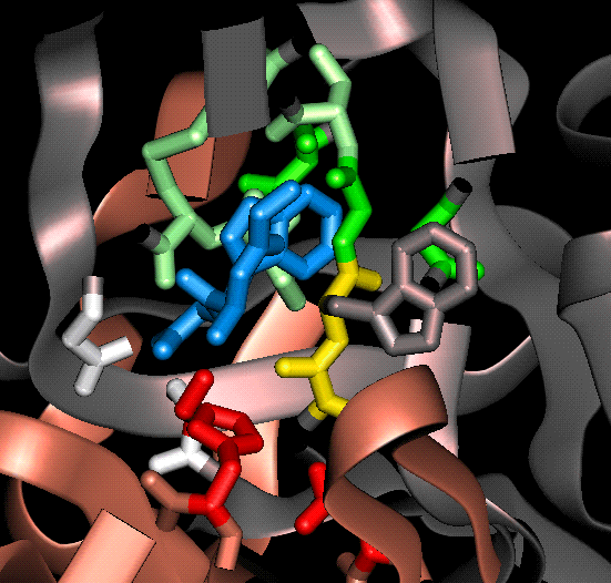
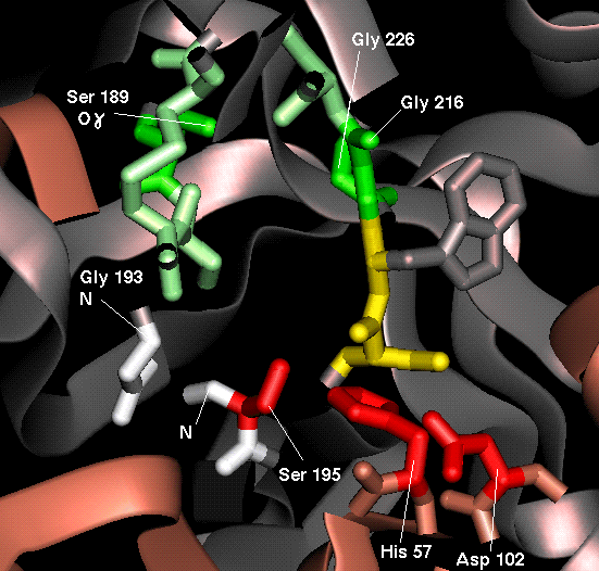
Catalytic Triad His-57 side chain 1 3,4  red
Asp-102 side chain 1 5,6
Ser-195 side chain 2 3,4
Oxyanion Hole Gly-193 main chain 2 3,4
red
Asp-102 side chain 1 5,6
Ser-195 side chain 2 3,4
Oxyanion Hole Gly-193 main chain 2 3,4  white
Ser-195 main chain 2 3,4
Specificity Pocket Ser-189 side chain 2 5,6
white
Ser-195 main chain 2 3,4
Specificity Pocket Ser-189 side chain 2 5,6  green
Gly-216 side chain 2 5,6
Gly-226 side chain 2 3,4
Main Chain Ser-214 main chain 2 5,6
green
Gly-216 side chain 2 5,6
Gly-226 side chain 2 3,4
Main Chain Ser-214 main chain 2 5,6  yellow
Substrate Binding Trp-215 main chain 2 5,6
Gly-216 main chain 2 5,6
Tripeptide cleavage
yellow
Substrate Binding Trp-215 main chain 2 5,6
Gly-216 main chain 2 5,6
Tripeptide cleavage  blue
product
blue
product
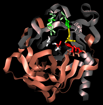
The following diagrams are of chymotrypsin complexed with a tripeptide (crystal structure 8gch). This tripeptide (Gly-Ala-Trp) is in fact a product of autolysis, i.e. a peptide fragment produced by the hydrolysis of chymotrypsin by another chymotrypsin molecule; refer to Part 1 .
(Crystallography note: numerous different types of peptide fragment occurred in different chymotrypsin molecules in the protein crystal; this tripeptide results from the overlapping electron density of different asymmetric units.)
Views are shown both with and without the tripeptide fragment.
A view looking into the substrate-specificity pocket, without tripeptide:
The same view with tripeptide:
 a different view to illustrate
the main-chain hydrogen bonding to the substrate/product
a different view to illustrate
the main-chain hydrogen bonding to the substrate/product

Back to Main PPS Index
J. Walshaw
 close up
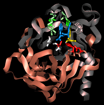
 close up