Haemoglobin belongs to a family of haem-proteins which contain a haeme prosthetic group. The best known haeme is Fe-protoporphyrin IX. Other members of the family include: b-type and c-type cytochromes, catalases and peroxidases. The function of Hb is to carry oxygen to cells, and it has to do so keeping the Fe++ in the reduced state.
Hemoglobin is a tetramer consisting of 2 pairs of identical dimers, alpha1beta1 and alpha2beta2 subunits. Each of the 4 chains contains one haeme group, in which the Fe ion is coordinated to the 4 nitrogen of the tetrapyrrole ring and the nitrogen of His87 of helix F. The sixth coordination site is free for ligating oxygen.
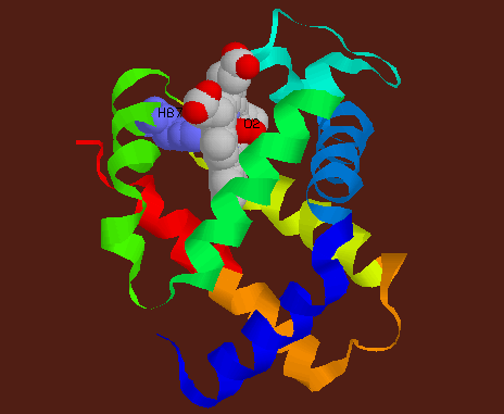
![]() Single
alpha chain of human HbA showing the 8 helices (from blue to red),
Haeme in CPK colouring with O2(red) and His87.
Single
alpha chain of human HbA showing the 8 helices (from blue to red),
Haeme in CPK colouring with O2(red) and His87.
Tetrameric Hb occurs in two conformations: the oxygenated T-state and deoxygenated R-state. The individual chains within each dimer are tightly packed together, but the two subunits are reversibly held together by a multitude of salt bridges and hydrogen bonds in the deoxy state.
Hb is the prototype of allosteric proteins. When oxygen binds to deoxyHb, the haeme ring slightly flattens, causing one helix (EF turn, F helix, FG turn) to move. This destabilizes the salt bridges between this helix and a helix on another subunit. A new inter-subunit salt bridge configuration is formed. This switch of configurations at the subunit interface causes the alpha1beta1 and alpha2beta2 dimers to rotate 150 and translate 0.8 Angstroms relative to the each other. The allosteric movements increase the O2-affinity of haeme in other chains, giving rise to cooperativity.
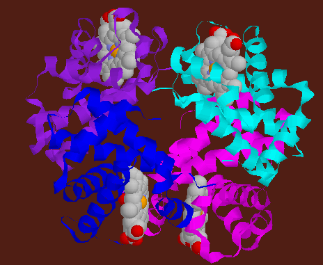
![]() Human
deoxyHbA, alpha above beta units. alpha1(cyan), alpha2(blue), beta1(magenta,
beta2(purple).
Human
deoxyHbA, alpha above beta units. alpha1(cyan), alpha2(blue), beta1(magenta,
beta2(purple).
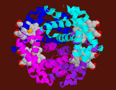
![]() Human
oxyHbA, alpha above beta units. alpha1(cyan), alpha2(blue), beta1(magenta,
beta2(purple).
Human
oxyHbA, alpha above beta units. alpha1(cyan), alpha2(blue), beta1(magenta,
beta2(purple).
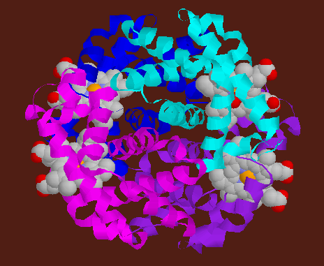
![]() Human
deoxyHbA, one alpha/beta dimer above the other. alpha1(cyan), alpha2(blue),
beta1(magenta, beta2(purple).
Human
deoxyHbA, one alpha/beta dimer above the other. alpha1(cyan), alpha2(blue),
beta1(magenta, beta2(purple).
![]() Human
oxyHbA, one alpha/beta dimer above the other. alpha1(cyan), alpha2(blue),
beta1(magenta, beta2(purple).
Human
oxyHbA, one alpha/beta dimer above the other. alpha1(cyan), alpha2(blue),
beta1(magenta, beta2(purple).