 Structure and Function of Green Fluorescent Protein
About this project
These webpages about Green Fluorescent Protein (GFP) have been developped as
a dissertation project for the
Principles of Protein
Structure Using the Internet course, in which I have participated as a
student. You may also want to have a look at my colleagues'
projects.
At this point I would like to thank all the tutors, consultants and fellow
students,
who made this course work - and it has often been great fun, too!
I am also indebted to Prof.
George Phillips, who has kindly given me the coordinate file of GFP prior
to its PDB release. Finally, I am grateful to Horst-Joachim Schirra for his
help with corel-draw and for patiently discussing many ideas of this project
despite his own heavy workload.
The project is best viewed with Netscape2, but apart from a few messy sub- and
superscripts Netscape1 should be okay as well.
Feel free to mail me
your comments!
Abstract
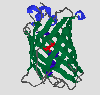
GFP is an extraordinary protein in many respects: It is fluorescent and its
fluorophore is made up of modified amino acid residues. Moreover it is the first known
example of a Förster cycle within the core of a protein. Furthermore its
crystal structure has recently been solved and the protein turned out to have
a new structural motif, called the beta-can. On the following pages I will set
out to discuss some of the most interesting features of this unique protein,
starting out with its function in the introduction, followed by a view on the
more or less isolated chromophore and finally presenting the three-dimensional
structure of this fascinating protein.
Content
Introduction
- The in vivo role of GFP
- Some basic properties of A. victoria GFP
The Chromophore of GFP
- Chemical Structure of the Chromophore
- Biosynthesis of the Chromophore
- Excitation and Fluorescence Emission Spectra of GFP
- Förster cycle
- Mutations affecting the of Tyr66
- Mutations of the chromophore forming residues
The 3D Structure of GFP
- The Beta-Can Structure
- Topology of Folding
- Cysteins
- The Environment of the Fluorophor
- Tryptophan Fluorescence
References
 [Introduction]
 Silke Jonda's PPS2 project
Structure and Function of GFP
updated 28.11.96