3.0 Types of Secondary Structure
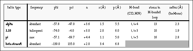
n - the number of residues per helical turn
r - the rise per helical residue
By convention, a positive value of n denotes a right-handed helix. (Curling the fingers of your right hand along the helical path, your thumb will point in the direction of your fingertips if the helix is right-handed.)
Table I: Parameters for common proteins helices. Values are given for pure geometrical forms of these conformations. Here n is the number of residues per helical turn, r is the helical rise per residue, and p is the helical pitch (Å/turn).
Historically (Brag, Kendrew & Perutz, 1950), helices were often designated by the number of residues per helical turn and the number of atoms in one hydrogen-bonded ring (as a subscript or following a decimal point but here due to formatting limitations of html, I will have to use parenthesis). Thus the alpha, 3.10, and pi helices were designated 3.6(13), 3.0(10), and 4.4(16), respectively. This nomenclature persists only for the 3.10 helix since it can lead to ambiguities (e.g. 3.6(13) and 3.7(13) are both alpha helices).
A word of caution in viewing 2-dimensional stereo pictures: There exist two ways of viewing stereo pictures 1.) crosseye and 2.) walleye. To be stereochemically accurate, stereo pictures need to be produced and viewed with the same convention. If not, the resulting picture still appears in stereo, but the stereochemical designations are reversed.
Helices are the most abundant form of secondary structure containing approximately 32-38% of the residues in globular proteins (Kabsch and Sander, 1983). Of all possible geometric forms, the alpha helix is by far the most abundant. Short segments of 3.10 helix are found. As we shall see, ends of alpha helices can contain a single 3.10- or pi-like hydrogen bond (3.1.4.1). The gamma helix was also predicted to be a possible structural element in proteins by Pauling and Corey. This helix has the designation 3.6(14) and was never observed. It may be an interesting exercise to create this very different helical structure (pencil and paper are sufficient if models are not at hand), once you have had a chance to look over this section. You will discover (if you had not already) two very different families of possible helices from polyamino acids which differ in the number of residues in the hydrogen-bonded loop: (3n + 4) and (3n + 5).
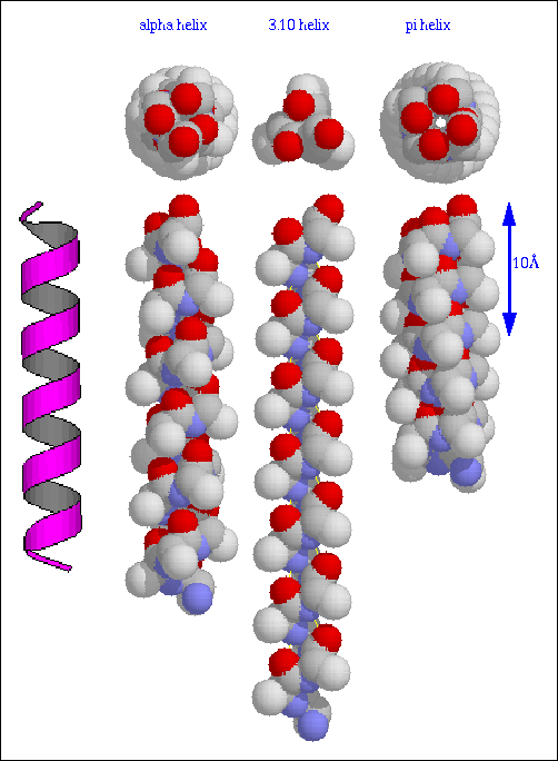
Figure 2. Three regular polypeptide helices. A cartoon of a icosapeptide in a right-handed alpha-helical conformation is shown on the far left. Idealized model alpha-helical (phi = -57.8, psi = -47.0), 3.10-helical (-74.0, -4.0), and pi-helical (-57.1, -69.7) coformations of a polyalanine icosapeptide are displayed (without hydrogens) using CPK representations. Views are perpendicular to the helical axis with the N-terminal at the bottom (lower) and following a 90 rotation (top). Standard CPK color is used with the exception that the alanine side chain (CB) carbon is a lighter shade of grey for distinction from backbone carbons. All structures are reproduced at the same scale using the program RasMol.
Generated with CERN WebMaker
No Title - 31 MAY 96
written by Kurt D. Berndt